
3D printing is not only an alternative to established production methods but also allows the creation of completely new product classes. One of these novel applications is in the field of medicine and science. In medicine, 3D models can make anatomy and pathology literally tangible. In science, the applications are very diverse. On one hand, custom-made objects for experimental setups can be produced using 3D printing. On the other hand, microscopic or even submicroscopic structures can be greatly enlarged and represented as physical models. Typical applications include anatomical models for surgical preparation, models for education, and models for laboratory and experimental needs.
This article provides an overview of the most important application areas in education, science, and medicine.
Anatomical models for surgical preparation
Through 3D printing, anatomical models can be represented in a patient-specific manner. In some cases, complex situations arise where an individual physical model provides a better representation of the condition than a purely digital model.
By simulating the realistic situation, the surgical steps of the procedure can be more accurately planned, reducing the duration of the operation and the patient’s burden. Therefore, the production of anatomical 3D models is a suitable method to give trainees and experienced physicians an overview of complex and extraordinary situations. Typical situations where anatomical models are applied include congenital heart defects in newborns with unclear or complex anomalies, severe fractures, or complex tumor removals.

For an accurate reproduction of the target structures, a CT or MRI scan can serve as a basis, which is usually available in the DICOM format. DICOM is an open standard for a collection of medical images and the transfer of image-related data.
You can get an impression of DICOM data, for example, on the Dicom Library platform, where DICOM datasets are published for scientific purposes. To design a 3D printed model from the scan, the file needs to be exported into a suitable 3D format. 3D data in the STL format is created based on the different grayscale values using an algorithm. Manual adjustments are then made to complete the model, such as removing artifacts and refining important details. Once the data is completed, the model is ready for printing.
Three additive technologies are particularly used for printing anatomical models:
- Polyjet: Allows for a two-component print with transparent & opaque or solid & flexible materials. The former is used especially to visualize delicate structures in spatial context, while soft materials are used to mimic the tactile properties of tissues.
- Lasersintering / HP Multi Jet Fusion: These processes use polyamide and work without support structures. The material is very stable and is often used when, for example, bone structures need to be drilled for testing, complex structures need to be depicted, or stability is a crucial factor. Since 2019, the MJF process also allows for color printing.
- Colorjet: This plaster-like material is suitable for simulating the tactile properties of bones and representing full-color structures (e.g., to distinguish different areas). However, the material is fragile, so the models should not be subjected to large forces.
Models for education
Models for education are usually very specific. Commercially available models are often highly simplified and limited in selection, so specific models need to be created through elaborate work.
3D printing can provide a faster and more cost-effective alternative in these cases. 3D models are commonly used in anatomy, biology, chemistry, and geology. In these various fields, three-dimensional visualizations help understand and comprehend the structures and functional relationships.

In anatomy, organ models are mainly created. 3D printing offers several advantages over commercially available standard models:
- Pathological changes can be individually depicted
- Cross-sections can be placed arbitrarily
- The print can be done at a very high three-dimensional resolution, with the models often being much more detailed than conventionally produced models
The basis for this is usually data from medical imaging procedures (CT, MRI) of patients or, in some cases, from specimens. Less frequently, 3D scans of plastinates or similar models are used.
In biology, 3D printing is used to represent microscopic or sub-microscopic structures in an enlarged scale as plastic models. It doesn’t matter what the structures are, as they can range from complete model organisms to isolated cell structures. The basis for this is usually high-resolution imaging techniques such as X-ray microscopy, etc.

In chemistry, biochemistry, and molecular biology, among other things, molecular models or interactions can be represented. More information can be found here:
- Applications and general information about protein and molecule models
- Creating a 3D-printable protein model from a Protein Data Bank (PDB) file.
Depending on the specific application, finding an appropriate file for the models can vary greatly. In some cases, 3D data is already available, such as in the case of molecular models or 3D X-ray microscopy images (as in the example of the fly depicted above). In other cases, there is no existing 3D data and it needs to be created. This can be done through a 3D scan or manual design based on existing images or sketches.
Laboratory and Experimental Needs
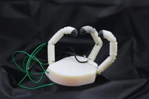
All experiments have one thing in common: they venture into uncharted territory. Therefore, it is often necessary to develop individually designed laboratory equipment or experimental tools. Traditional model building can be very time-consuming. With 3D printing, comparable results can often be produced in a few days. The range in this area is very wide, from smaller objects such as custom grippers to complete experimental setups for complex experiments. 3D printing is a suitable manufacturing method. Since very specific materials or surface properties are often required, which cannot be achieved with additive manufacturing processes alone, the experimental setups are often complemented by additional parts from other production methods.
We are the partner for research institutions, university hospitals and universities
As a 3D printing service provider, we are a recognized and established partner of academic institutions. We have experience in working with over 20 universities, numerous hospitals and many research institutions. Academic institutions of public sponsorship enjoy advantages with us such as payment on account.
About 3Faktur: 3Faktur specializes in 3D printing, rapid prototyping, and rapid manufacturing. We utilize HP’s Multi Jet Fusion technology and offer various materials for prototyping and series production. If you have any questions about your project, feel free to contact us.
